When Women Birthed Mooncalves and Moles
The Display of Fetal Remains and the Invisibility of Females in Museums
DOI: https://doi.org/10.52537/humanimalia.10030
Emily F. Porth is a PhD Candidate and sessional lecturer in the Faculty of Environmental Studies at York University in Toronto, Canada. She is a feminist anthropologist and animal advocate whose interdisciplinary research focuses on how the visual narratives in museum displays can be reimagined to encourage relationships of respect between humans, and between humans and other species. She is currently completing her dissertation, titled How to See Differently: Social Inclusivity and the Display of Bodies in British Museums.
Email: e.porth@surrey.ac.uk
Humanimalia 4.1 (Fall 2012)
Abstract
The Hunterian Museum, part of the Royal College of Surgeons in London, England, is unique in that its displays are focused on the anatomical collections of 18th century surgeon John Hunter, and today the museum is designed to exhibit Hunter’s collections in the way he would have at that time. As such, humans and animals are unusually displayed in the same galleries, and this includes a large number of wet-preserved human and animal fetal bodies. Significant work has been done within feminist studies of science to examine the politics around the icon and visibility of the human fetus, and on how the fetus has become the primary actor in the origin stories of contemporary Western society. However, the critical investigation of animal fetal bodies and their role in this origin story appears absent altogether, and little work has been carried out on how foetal display in museums essentially renders females of all species invisible.
An Introduction to the Hunterian Museum
I passed through the regal Roman columns that grace the front entrance of the Royal College of Surgeons in London, England and collected my dark yellow “Museum Visitor” badge from a security guard at the front desk. Upon walking up the stairs to the second floor and then entering the museum, I was greeted by a volunteer working at the desk to the right, who directed me toward the large laminated museum guides located under a bust of the museum’s namesake, John Hunter.
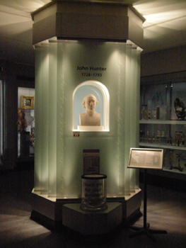
The Bust of John Hunter (Photo by Emily F. Porth)
The light is kept quite dim, owing to the fragility of the many wet-preserved specimens in the main part of the museum: row upon row of bits, sections, and entire human and animal specimens. They were delicately dissected and suspended in an alcoholic solution in the interest of their long-term preservation, most having been prepared by Hunter himself in the latter half of the eighteenth century, and the quality of the specimens is immaculate. The museum collection also includes surgical instruments, skeletal remains, wax and plastic models, as well as a few odd specimens that have been preserved from the ravages of time through the old technique of rapid dehydration, followed by the application of a thick coat of lacquer. It is the spirit collections however, in their seemingly endless rows of glass jars of innumerable size and content, that dominate this space on the translucent shelves and give it its name: “The Crystal Gallery.”
The most imposing sight on entering the main gallery is the skeleton of “The Irish Giant,” a man named Charles Byrne who exhibited himself in life as the world’s tallest man, and who ended up — against his explicit wishes — as part of John Hunter’s expansive collection. Now, as an iconic figure associated with the museum, it is incredibly unlikely that his remains will be removed from display, even though the museum and the public are well aware that he did not wish to be displayed in death as he was in life.
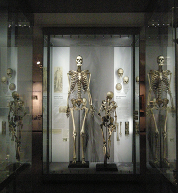
“The Irish Giant” (Photo by Emily F. Porth)
The Hunterian Museum is a medical museum and, in the tradition of such museums — and of science itself — its specimens come from people and animals who frequently remain unknown and anonymous in the collection catalogue, and who are almost always displayed in the same fashion: without names, and without individual histories. The skeleton of Mr. Byrne is one of few specimens that has a documented story, and whose story is told within the exhibitions.
Passing through the galleries, I was struck by one thing after another; the museum is an almost overwhelming visual cornucopia of beings, and pieces of beings, that represent the diversity of life on this planet, as well as the incredible progress made in the eighteenth century in understanding anatomy and the workings of biology. It is also a strong indicator of the ambition and curiosity that drove Hunter’s medical research and collecting. Some specimens, such as a piece of honeycomb, or various insects, seem more innocuous than others that, frankly, left me unsettled. The more shocking beings seem to appear more sharply in the glass shelves: a small pregnant water vole (specimen 3462) is suspended upside-down in clear fluid, the skin over her bulging belly removed entirely to reveal numerous uteri that are connected together and curve like a large intestine outside of her body. On the other side of the shelves, part of a child’s face (specimen P 334) is preserved, riddled with smallpox sores that have burst and ulcerated. The face, unidentified as either male or female, is sawed across at the rim of the eyes with lower eyelashes left intact, and then also cut vertically midway down the side of the face, leaving the teeth in the mouth, but with an empty void behind it. The dye injected into the skin to color the sores left the flesh looking blanched but remarkably lifelike. By the time I had made it through most of the collection, I felt thoroughly desensitized to what I was seeing — and it seems likely that is part of the logic for locating the mammalian embryological and fetal specimens in the final corridor of the museum.
Particular biological similarities make humans feel emotionally closer to some other species. Erica Fudge asserts that the characteristics of big eyes and long eyelashes, for instance, compel consumers to feel more strongly about the plight of veal calves than chickens in factory farming. This is because we see the calves, in a way, as a mirrored version of ourselves, which promotes a very specific type of recognizability between human and animal (Fudge 40). In contrast, animals like fishes look so completely different from humans, that empathy is often more challenging in practice. At the Hunterian, there are fetal bodies representing the varying stages of development of multiple species — insects, reptiles, almost any type of animal you can think of — but it is those species that are most closely related to human, as well as humans themselves, that are grouped together near the end of the path visitors take through the museum
***
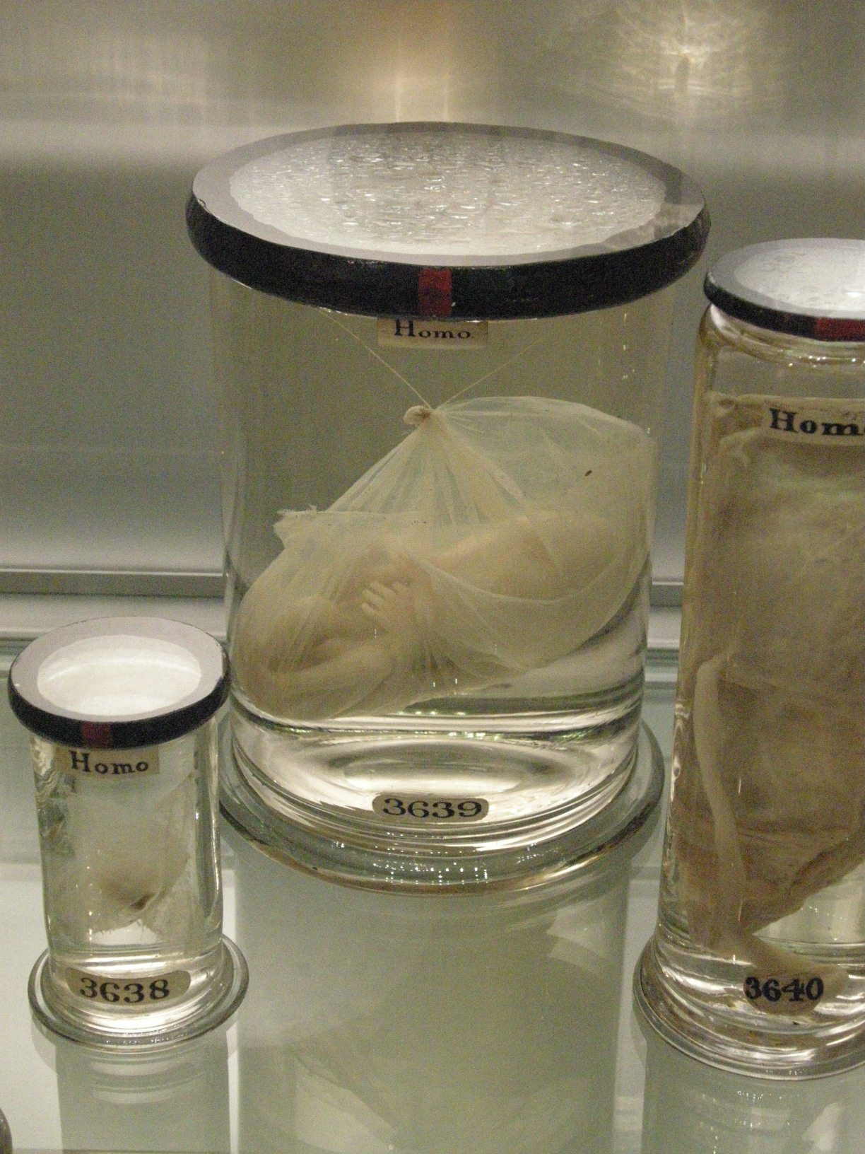
She sits in a glass jar labelled “3773”. Her skin is a waxy beige, lightened by the formaldehyde and alcohol that preserve her body, and she is completely hairless. It seems apparent that her eyes were not developed enough to have ever seen, had she been outside of her mother at that stage of development — it just would have been too early for survival. Her mouth is slightly open and teeth and tongue are apparent just inside of her prised lips. A large scar marks the length of her trunk, stretching from the groin, all the way up to what remains a large, gaping hole in the centre of her chest. Affixed in anatomical position, her arms rest in front, palms facing downward, and she hangs suspended, appearing almost to sit within the glass jar that has been her final resting place from sometime between 1760 to 1793. It is a protective glass womb to replace the pulsing, fleshy one she left behind. This one does not nourish the way her mother’s did, but with the correct proportion of chemicals within, it does sustain its own type of “life” for the body within it: an eternal one. Where did she come from? Under what circumstances was she removed from the female who nurtured her biological development? And if this was her fate, what happened to her mother?
Fetal Remains in the Museum
The catalogue entry from 1970 for the wet-preserved fetus described above is brief: “A female mammary fetus of a Kangaroo (Macropus sp.), at a slightly more advanced stage of development. In the centre of the abdomen is the mouth of the marsupial pouch. The lateral fusion of the lips is still maintained although it is in process of breaking down” (Dobson).1 One of the volunteers at the museum told me that she and the other specimens on this shelf were the first kangaroos that had ever been seen in London (personal communication, June 8/10), although this information doesn’t appear in any of the catalogue information about the kangaroo specimens. Did you initially assume the fetus I describe is human? If so, did you experience an emotional reaction to her? If you had known she was a kangaroo fetus, do you think your reaction would have been different? Would you have thought about where she came from and wondered about the details of her individual family history?
In this paper I argue that narrating the embryological origin story through traditional scientific display techniques has the potential to reinforce the systemic marginalization and devaluation of females of all species. When fetal bodies are exhibited without reference to the context of their acquisition, females become the “absent referent” and can essentially be morally abandoned as “incubators.” The purpose of this paper is to draw attention to the displays of bodies in scientific institutions that are presented, and perceived by most of people who visit them, as “objective” and “value-neutral.” Fetal remains occupy a sensitive space for some people because of their visual association with contemporary debates about whether or not a woman should be able to choose to terminate a pregnancy and, regardless of politics, the fetal remains at the Hunterian Museum are visually arresting bodies that attract a lot of attention from visitors.
During the summer of 2010, I carried out fifteen interviews with visitors to the Hunterian Museum.2 Nine of these interviewees voiced their interest or concern about the circumstances surrounding the acquisition of the human fetal bodies because no context was provided in the display about how they came to be in John Hunter’s collection.3 A woman I interviewed, who had recently had a baby herself, said, “the fetuses seem more brutal — it’s the idea that they were ripped out of somebody” (Penny, 16/06/10). Her frank words made an impression on me, and left me to wonder about the women who were the biological mothers of these fetal remains and essentially rendered invisible through the process of museum collection and display. It is also intriguing that interviewees so seldom used the word “mother,” or even “woman,” to describe the source of the human fetal bodies.
Of equal interest is my interviewees’ general lack of curiosity about the source of the fetal remains of nonhuman animals. They, too, are the offspring of mothers who have become invisible, and the presence of these fetuses in the museum is likewise generally unexplained in the catalogue and displays. My interviews reinforced the importance of drawing attention to the display of fetal remains because they are symbolic of the females, both human and nonhuman animal, who are not commonly given adequate representation in places of informal education, and who are in fact sometimes dramatically misrepresented, as I discuss later. I could have examined how other types of discrimination, including racism, classism, ableism, and heterosexism are apparent in the ways fetal bodies are displayed, but given the length of this paper, I have chosen to focus my analysis on females and the sexism that continues to exist within museum displays.
The “embryological origin story” is a concept developed by Lynn M. Morgan, an anthropologist whose book Icons of Life: A Cultural History of Human Embryos (2009) emerged as central to my work. She defines origin stories very basically as narratives that “are patterned, predictable accounts of how ‘we’ (the people) came to be” (8). Origin stories come from many different sources and are traditionally associated with religious beliefs, but embryology is the origin story of modern science — itself a system of belief — in which the embryo/fetus4 plays the key role, and it is the one that many humans take for granted as the “truth” for all people, regardless of religious or cultural beliefs, about the origins of all animals. Morgan poignantly remarks that, “the cultural ideology that portrays embryos and fetuses as natural, anonymous, free-floating creatures carries serious consequences” (138), and although her work deals solely with humans, my research at the Hunterian draws attention to how some of these concerns affect females of all species.
It is only possible to understand the embryological worldview by understanding what ideas about female bodies, pregnancy, and the unborn existed in the past, before this perspective became widely understood as “fact.” After situating my research within the historical context of the Hunterian Museum, I discuss changing perceptions in the West of what we today call “fetuses,” from the eighteenth century and through to the present, in conjunction with a discussion about the shifting ideas around how humans were connected to other animals. Duden (1999) in particular sheds significant light on ideas about human reproduction in the 1700s as a feminist historian of science. Her work on the case books of an eighteenth century German physician who wrote about the medical histories of nearly two thousand women has been instrumental in demonstrating that the ways we describe and understand the body are inherently cultural. Interwoven in this “herstory” are threads about religion and science, and how these shifting ideas about the origin story of humanity resulted in the changing relationships amongst humans, as well as with other beings.
These relationships continued to shift once ideas about evolution and natural selection became widely accepted, and embryology became an established field of study. Even today, museum exhibitions about evolution will include models of several animals, including humans, in their embryological state. These models are displayed together in a line to demonstrate how developmentally similar all species are, and how we share a common ancestor. I outline the defining features of the present-day embryological origin story, as Morgan has established them, and then discuss in more depth how culture is embedded within the scientific paradigm, of which the embryological worldview is a product.
Next, by bringing together the voices of interviewees with an analysis of the fetal displays in the Hunterian Museum, I explore how the gradual and largely uncritical acceptance of the embryological origin story has contributed to the exclusion and marginalization of both human and nonhuman animal females in the West. I go beyond discussing how fetal remains are displayed to analysing how they are narrated, and I specifically question whether or not these bodies should be situated within a context. Locating each individual specimen in its own “origin story” would render its mother visible and make females once again part of the story of reproduction, rather than abandoning them as the “absent referent.” It would also re-situate reproduction firmly within the social contexts with which it is inextricably entwined.
I am not advocating that fetal displays be removed from museums.5 However, in order to promote the increased inclusivity of females in the visual narrative of museum displays, I conclude by exploring some alternative display and interpretation techniques. This is possible even within museums like the Hunterian which present their collections within a historical context and need to remain mindful of the issue of authenticity.
Bodies on Display and the Hunterian Museum
How does the display of particular specimens, and the ways they are exhibited, reflect what humans think about themselves and each other? How does it mirror how humans see themselves in connection with other beings? In a museum, the assemblage of objects and specimens into an exhibition produces a “visual narrative”; as such, “collections as a whole, and also individual exhibitions, [can be considered] the result of purposeful activities which are informed by ideas about what is significant and what is not” (Hooper-Greenhill 3). Museum displays reflect not only unquestioned beliefs about relationships between humans and the rest of the world, but they also reinforce them. As such, it is essential to question the stories they tell visitors, and whether this narrative has the potential to be harmful and exclusionary. Answering these questions necessitates understanding the stories humans in the West learn and take for granted about their biological origins.
From the nineteenth century and into the present, human remains, as well as objects made and modified by humans, have usually been sequestered in “cultural history” museums,6 whereas animal remains have been displayed exclusively in “natural history” museums — two spheres that are, by their fundamentally artificial separation, represented as having little influence on each other and established in apparent opposition. It is thus a rare thing to see human and non-human animals displayed together in the same galleries as they are at the Hunterian Museum. It is done this way because specimens are displayed within what attempts to be an authentic historical context, and the present museum aims to display Hunter’s collection as he would have. According to Dr. Simon Chaplin, who was Senior Curator at the Hunterian Museum when it underwent the renovation that transformed it into the Crystal Gallery, “That was Hunter’s collection, with humans and animals displayed together; he was trying to show comparisons between different types of living bodies… Hunter wasn’t reducing humans, but putting them together as living things in the sense that it is not possible to understand one without the other” (Chaplin, 10/08/10). Chaplin remarked that the big idea in the museum’s renovation was to demonstrate that medicine in the eighteenth century did not have the same shape that it does now, and to highlight the importance of dissection in that period and in Hunter’s work.
When John Hunter passed away in 1793, he left an astounding collection of 13,687 prepared biological specimens (Asma 55). For Chaplin, however, the collection is fundamentally not just about dissection, but about presenting specimens for learning, and that was also Hunter’s intent when he began collecting, preparing, and displaying these specimens. While the Crystal Gallery is oriented “to create the visual spectacle of seeing thousands of specimens all together – to convey the same sense of awe and wonder that visitors would have experienced in the eighteenth century” (Chaplin, 10/08/10), the Hunterian Museum has been used for the education of surgeons and other scientists from its inception, and it now also educates a much wider audience since it was opened to the general public in 1995 (Asma 57).
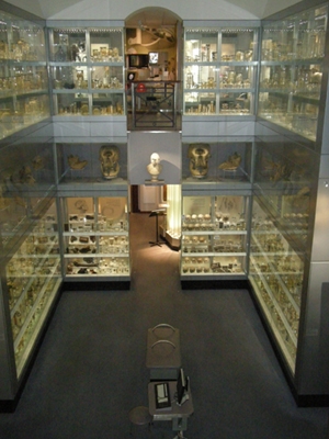
The Crystal Gallery (Photo by Emily F. Porth)
Fetal bodies have been preserved and displayed since people began collecting anatomical specimens. However, many science museums that previously had fetal collections on display have gradually phased them out, making the Hunterian and other anatomical museums unique spaces where they are still visible to the public. Given that the Hunterian Museum is focused on education, this makes it especially important to analyse the visual narrative that is told through the ways these bodies are displayed, and to question how it influences what visitors are learning.
Of Moles and Mooncalves: Human Origin Stories in 18th to 19th Century Europe
The fetus, as Western society identifies it today, did not exist for the women from whom John Hunter collected the fetal specimens that visitors can still see at the museum. In the eighteenth century, a woman was not acknowledged to be pregnant until she felt the “quickening” — when the developing child begins to move, at about eighteen to twenty weeks’ gestation (Duden 16). Even when a woman perceived herself as being “with child,” she did not envision a “fetus” or the stages of “fetal growth,” nor was there any guarantee that her state would result in a baby (Duden 13). For women in the eighteenth century, pregnancies could be “true” or “false,” and when a woman was “truly” pregnant, she would eventually give birth to a living child. However, as the German physician Wilhelm Gottfried von Ploucquet wrote in 1788, “Not everything that comes from the birth parts of a woman is a human being” (quoted in Duden 13). In the case of “false” pregnancies, “her swelling belly does not cover a yet unborn child, but a ‘something’ she conceived and will bring forth that is not recognised as a ‘child’, because it does not have a ‘human form’”(Duden 13).
Before the quickening, a woman whose menses had ceased was in a liminal state of being:
maybe she was “with child”, maybe not. Perhaps the cessation of her ‘monthly’ was due to some blockage, some “retention of menses”. What we today perceive as an abortion, a “miscarriage”, or the premature birth of a fetus, then, in the eighteenth century, could be perceived as emitting bad blood, the birth of a mole, a moon-calf, a “cleansing” of the womb, or as healthy flux against unhealthy stoppage. (Duden 16)
What is a mole or a moon-calf/moon-child? From the time of Aristotle, it was thought that “in the first weeks after conception, nature likes to shelter all kinds of beings, including little children, moles, monsters” (Duden 17). In the words of a physician named Johann Storch, who practiced medicine in Eisenach, Germany in 1725:
“a mole is a growth which, after carnal mingling, has been conceived in the womb, nourished through the generative power, and kept as a misshapen thing for a while, until nature expels it as something useless, with the same movements she uses for real birth”... [they are] useless beings, stagnations, which “nature seeks to expel from the body”. (qtd. in Duden 17-18.)
Essentially, until the middle of the eighteenth century, what we now understand to be a fetus was interpreted by anatomists as “a big-headed monster, a mooncalf, a misshapen thing” (Duden 20). What significance does this have for our understanding of human-animal relationships, if human females could birth other particular types of animals, albeit “useless” ones?
As evidenced from Duden’s case study, a belief in moles and moon-calves as part of human reproduction was not just part of a vibrant folk religion or local beliefs, but was part of official medical discourse across most of Europe. The fetus, as we know and understand it today, was simply not a part of this discourse: until the middle of the eighteenth century, illustrations of a human baby in utero usually resembled a fully formed small man who was often depicted sleeping, or doing some rather acrobatic activities. Over time, however, visualisations of the unborn began to look more like an infant, and details about the womb with the umbilical cord, etc. were added (Newman 28-35). However, knowledge about obstetrics and human development changed dramatically over the course of the next one hundred years as the embryological origin story developed, and with it ideas about the relationship between humans and other animals were altered as well.
There were many shifts in thought across Western Europe during the 1700s and into the 1800s about what was then called (and is still labelled as such in the Hunterian Museum) “products of generation.” It was not until 1782 that there is any record of change in term from “generation” to “reproduction”; this term seems to have been introduced by the natural historian and scientist Georges-Louis Leclerc, Comte de Buffon (1707-1788). “Reproduction” had previously only been used in reference to natural history, anatomy, or scientific contexts (Jordanova 372). This shift in terminology was met with some hostility, as Jordanova notes that the term “reproduction” had the implication of bringing humans to the same level as other living beings, including plants: “The fear of levelling was widespread in the period, prompted as much by political radicalism as by ideas that denied the specialness of ‘man’ or the presence of the immortal soul” (Jordanova 372).
During the medieval period (roughly 400 – 1500 AD in Britain), science was undertaken for the sake of religion — from the Christian Church’s perspective, nature contained patterns, but it was ruled from above by God, and so had no spirit or will of its own. At this time, a sharper line of division developed between humans and nonhuman animals, and because only men had been made in God’s image, humans (and males, in particular) were perceived to have been given dominion over all others (Noske 45). By the sixteenth to eighteenth centuries, dramatic change was underway in Western Europe. This included: colonial expansion; the weakening of Christian unity under papacy; the discovery and use of the scientific method; the proliferation of industrialism; and the rise of nationalism, individualism, and mercantilism across the continent (Kinsley 111). The Bible and Christianity were used to justify and reinforce many of these changes, particularly scientific pursuits and colonial domination, which were directly tied to the emergence of museums and the “cabinets of curiosity” that functioned as status symbols for the state and the wealthy.
Most of the prolific philosophers during this time were “working for God as well as science” (Coates 79). Nature could only become unimportant and subjected to the rigours of scientific scrutiny when humanity saw itself as separate and distinct from the natural world. Noske identifies the shift in this perspective as resulting primarily from the mechanical worldview solicited by Francis Bacon (1561-1626), René Descartes (1596-1650), and Sir Isaac Newton (1623-1747). These men embarked on a quest to discover nature’s “laws,” a process that created the scientific worldview and the idea that its findings represent Truth (Noske 53-54). Descartes, in particular, defined consciousness and thought as the final proof that humans were inherently separate from all other creatures on earth (Coates 17); “because consciousness could not originate in matter, only humans had a soul, which was given by God” (Preece 136). In this worldview, non-human animals are machines because they “lack mind” and, therefore, have no soul: an animal “lives in the world, responds to the world, but it cannot know the world. Any sign of knowledge that might be shown is mere instinct” (Fudge 99).
Once scientists became more interested in proving how different humans were from other animals, and more anatomical work was carried out on human cadavers from the early eighteenth century, the idea of a woman birthing moon-calves and moles became increasingly outdated amongst surgeons like Hunter who were actively collecting human and animal “products of generation.” Hunter and his older brother William, who was a prominent obstetrician and gynecologist, were likely at the head of this movement because of the research they generated through each of their extensive collections of human and animal specimens.7
The work of these scientists also led to different ways of viewing human female bodies, which have long been closely associated with animals and nature through their capacity to bear offspring. It was assumed that the “bodily demands” of women interfered with their ability to act independently and “rationally,” and thus put into question their capacity to be agents in their own right (Campbell et al. 5-6). In fact, part of the legacy of Descartes’ distinction between mind and body is “the equating of women, children, animals, and ‘the natural’ with one another and with the despised body,” a perspective that Elizabeth Spelman describes as “somatophobia” (Adams and Donovan 2). By the end of the eighteenth century, the idea of “woman” was universally disenfranchised, regardless of age, ancestry, or class, and natural historians proposed a “universal reproductive woman” (Schiebinger, Nature’s Body 6). Schiebinger attributes this significant interest in reproduction to a perceived need by colonial governments to increase populations in both the colonies and in Europe itself. Through the nineteenth century, “objective” scientific research was actively used to demonstrate the “natural” intellectual inferiority of the universal woman, as well as lower class Europeans, all non-European humans, and non-human animals. “Scientists, with their privileged knowledge of nature, became consecrated priests in a new secular order, intermediaries between the laws of nature and states” (9), and females of all species became valued primarily through their capacity to bear offspring. Whether wild or domestic, female nonhuman animals were prized for their ability to produce food for humans, and for their role in producing more animals that could be collected as new specimens in museums and zoological parks. For human females, their value lay in producing additional child or slave labor, or as the means to further populate Europe and the colonies with “desirable” people.
Although Hunter, his brother, and select other scientists collected and displayed fetal bodies as teaching tools for other medical professionals from the middle of the eighteenth century, it was not until 1799 that anatomist and physician Samuel ThomasSoemmering displayed a series of images of male and female fetuses lined up by age and size in a small booklet he published under the title of Icones Embryonum Humanorum, which translates simply as “Images of Human Embryos” (Duden 18). This strategy deviated from the historical norm of always depicting the developing child as male, but it has also served to reinforce the gender binary, which is still problematic. Soemmering notes that although he captured each fetus as an individual, it is still meant to act as a type that represents all fetuses at that point in gestation (Duden 22). This visualization technique echoes how scientists and curators will choose a single non-human animal in a museum collection to represent its entire species: this is called a holotype. It is the anonymity of specimens in museums that allows spectators to see them as a “type,” rather than as individuals, and it also gives visitors the right to look. The latter is particularly so when gazing at fetuses, which are marginalized in society outside of the pictures available through ultrasound technology. This opportunity is made possible only through the authenticity of the bodies, as well as the viewing permission that is granted through an exhibition (Desmond 371).
Through Soemmering’s booklet, the display of fetal bodies in their successive stages of development became standardized. It is an important indicator of the role science continues to play in demonstrating what appears to be the inevitable progress of humanity through the evolution of the individual human. Non-human animal fetuses were widely collected too (those from exotic species having the most prestige), but continue to be referred to as “comparative specimens.” By designating humans as the standard by which other animals are judged, humanity is represented as the pinnacle of evolution.
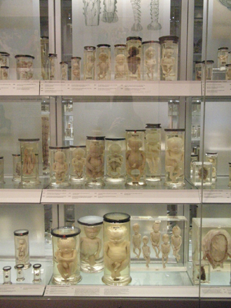
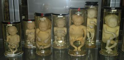
Stages of Fetal Development (Photo by Emily F. Porth)
The collection of human and non-human animal fetal bodies — catalogued and displayed without any reference to their mothers, families, or the context of their procurement — slowly began to change religious and folk origin stories, and human offspring that had previously been interpreted as moon-calves and moles were re-defined as human. Through this, the magical reproductive link to other species in the West was severed. Although it certainly cannot be claimed that animals were treated with more respect prior to this time, in general people had an undeniably closer relationship with the natural world and other animals because so many were based in rural areas and still profoundly dependent on the land for survival. Further, the new visualization of fetal bodies dramatically changed what it meant to be pregnant for women; both they and European society began to re-interpret women’s bodies and their “products of generation.” This marks the point at which fetuses came to be seen as individual entities separate from their mothers; as they were collected from females whose identities and circumstances were not only anonymized, but designated as completely unimportant by the emerging scientific community.
Animal Kinship: Darwin’s Theory of Evolution and Reproduction
Charles Darwin (1809-1882) spent quite a bit of time at the Hunterian Museum in the late 1830s to early 1840s as he and the curator, Richard Owen (1804-1892),8 inspected the fossils that Darwin had brought back from South America. Asma (2001) surmises that Hunter’s large collection of teratology specimens9 “served as the backdrop for Darwin’s and Owen’s early discussions of extinction. Individual bodies and organs, preserved in the act of fluctuation from the norm, were suggestive of flux at the species level,” and this is a preoccupation that Asma claims surfaces repeatedly in Darwin’s diaries from the period (66). When Darwin finally published On the Origin of Species in 1859, it was ground-breaking in part because it provided a scientifically feasible mechanism to explain the idea that species were not created separately, nor did they exist in fixed biological forms. Rather, they were each subject to processes of natural and sexual selection that, given variation, heredity, and long spans of time in which to act, could account for the vast diversity seen in the natural world (Noske 63). Perhaps most controversially, as Noske articulates, “the random character of natural selection turned living nature into an accidental object, brought about by mechanical forces and made to exist in a cold, meaningless universe” (66). Within this universe, God seemed to have no place in the new scientific narrative of earthly creation.10
While largely focusing on the evolutionary forces acting upon plants and animals, it was clear that Darwin included human beings within his theory, even though it was a point at which he only hinted near the end of On the Origin of Species: “Light will be thrown on the origin of man and his history” (Darwin 488). Evolutionary theories strongly implied a connection between human and animal life in a way that previous Christian accounts had not, and this was often emphasized through a comparison between human and animal embryonic development. However, because human embryos were very difficult to obtain, these developmental similarities were often applied last to humans, and so they remained ideologically distinct from other species (Morgan 49; 47).
The view that phylogeny recapitulated ontogeny, or that the embryological development of “higher” animals went through stages roughly corresponding to those of the lower animals from which they developed, was articulated and popularized by the German biologist Ernst Haeckel (1834-1919) in the middle of the nineteenth century (Morgan 49). Around this time it was discovered human embryos have a “tail” from the forty-first day after conception for a total of about two weeks, and although some of the public found this news blasphemous, others saw it as proof of recapitulation (164). Regardless, the embryonic tail became an important line of demarcation between humans and other animals. Further, the embryonic tail demonstrates how anatomical traits became imbued with symbolic meaning, which effectively helped to create what it meant to be an embryo (168).
Haeckel’s leading opponent, the anatomist Wilhelm His, Sr. (1831-1904), instead insisted that embryology needed to focus exclusively on human development, and he helped to shape the ensuing mania for collecting human embryos. During the latter half of the nineteenth century, scientists went to drastic lengths to obtain human embryos at the earliest possible stages of development (see Morgan). These specimens were then shipped to scientists seeking them through elaborate “supply networks” (Buklijas and Hopwood), which Morgan appropriately labels a “patriarchal project, as men circulated the most intimate contents of women’s bodies among an old boy’s network” (89). This is not unlike the collection practices of explorers and naturalists who feverishly “collected” (i.e., killed) almost every animal who crossed their path in the name of acquiring a “perfect” specimen. As there was a demand for comparative fetal specimens by scientists and curators as well, pregnant animals in the wild were also seen as valuable prey for colonial explorers.
One continues to see parallels in the treatment of female human and nonhuman animals during the early twentieth century, when embryo-harvesting techniques used on laboratory-raised rhesus macaques were employed as a model for finding human embryos in tissues from hysterectomies (Morgan 127). This collection process involved re-branding fetal remains from waste tissue to active tissue, and re-positioning all females as mechanistic “incubators,” rather than as mothers. This effect was achieved with human fetuses through popular literature that likened human and bird development: after all, if embryos were like eggs, they could be sorted, collected, and studied on their own merits as mass-produced specimens, without regard for the lives of the women involved (56-57).
Historically, and into the early twentieth century, women who miscarried or aborted pregnancies handled fetal remains privately, and if scientists asked for them, families seemed to have no ethical issues or personal attachment to the remains. That does not mean, however, they did not grieve for the emotional loss of a pregnancy — the tissue was simply valued differently. For instance, the remains of quintuplets are on display at the Hunterian Museum (specimen 3681): born premature at five months’ gestation, three babies were alive at birth and died shortly afterwards, and the other two were stillborn. In a rare case (probably due to their remarkability as specimens), the information about the quints’ origins is found in both the museum catalogue and on the display label:
They were born to a mother in Blackburn, Lancashire in 1786. A local surgeon called John Hull attended the birth… In his report Hull describes how he was allowed to take the bodies of the fetuses, but was not permitted to take the placenta which was burnt in accordance with local custom. He sent the preserved bodies to London and they were placed in John Hunter's museum. (RCSENG)11
Eventually, the collecting activities of embryologists modified women’s behavior, and women learned not to discard dead embryos and fetuses in their own way (i.e., burial on one’s property, or disposal down the privy). They also learned that the most acceptable course of action was to surrender dead embryos and fetuses to doctors (Morgan 82). This shift accompanied the more widespread medicalization and institutionalization of reproduction and birth, of humans as well as of other animals in the factory farm industry, zoological parks, and purebred companion species. By the early 1900s, images of embryonic development were commonplace across the West, “yet court cases and interviews reveal that for a long time working-class women seeking abortions still tended to speak, not of eggs, fertilization and embryos, but about needing to restore an interrupted flow and rid themselves of a waste material” (Buklijas and Hopwood). Clearly, this shift in viewing pregnancy, women’s bodies, and products of generation did not occur ubiquitously across all sectors within society, or simultaneously within all societies in the Western world. However, evolution and, by extension, embryology, did change how women saw their own bodies, pregnancy, and the process of reproduction. By the 1930s, the embryological origin story was widely accepted in the Western world by most sectors of society. Different religious and secular worldviews took knowledge of the fetus and made it their own (for instance, the question of when “life” begins and its influence on law and public policy regarding a woman’s right to terminate a pregnancy), but it was still considered to be a taken-for-granted biological reality, regardless of interpretation.
Although evolutionary theory made tangible the biological kinship that humans share with other animals, in practice it rarely changed the attitudes of humans toward other animals. And, as women and female animals have been further exploited through their reproductive capacities within the embryological worldview, primarily as incubators for new life, the fetus emerged as an independent agent and, potentially, as an individual capable of being ascribed personhood.
The Embryological Origin Story: The Contemporary Scientific Perspective
Morgan identifies embryos as the central actors in Western society’s modern origin story (4). As noted above, not all peoples believe or believed that the being developing in a female’s uterus is, in fact, an embryo or fetus. However, fetal development has become a taken-for-granted “reality” for most of us through the increasing visualization of the biological development of the fetus, particularly through ultrasound technology. That fetal bodies have the “capacity to become a sacred image of life itself is demonstrated by its ability to represent the human, the nation, the species, and the future” (Franklin 64).
Morgan identified several assumptions within the embryological origin story. First, this view of development assumes that the path from embryo, to fetus, to baby is the only way to become a full human being, and it supposes that all humans begin as embryos, and we were all fetuses at one point. Next, the embryological view assumes that the production of all human pregnancies is human embryos — they cannot come to bear another species, or hybrid of another species, or an inanimate object (11). Third, this view assumes that embryos are “amoral biological entities, defined and classified solely by their genetic and anatomical features,” which is harmful because it is a type of biological reductionism that does not take into consideration “the spiritual, moral, or social circumstances that generate embryos”(12). The final assumption in the embryological origin story is that it is the only story, that it is a story free of subjective belief, and that is true above all other stories. Over the past two generations, humans in the West have come to know this story as not as a cultural artifact, but rather as “the facts of life” (12).
The Culture in Science
Why is it critical to see our understandings of biological processes as cultural artifacts? In her book The Woman in the Body, Martin builds on the metaphor of baking a cake to illustrate the importance of critically analyzing popular discourses about female bodies:
I assume that women would not menstruate or give birth if they did not have physical bodies and if those bodies did not contain genes, hormones, or many other things. Questions about how genes or hormones function in human lives are legitimate, but even complete understanding of them could never settle any human matter. If we focused only on completely understanding the physical components of the cake, we would lose the person who baked it, the occasion it was baked for, and the people who were there to eat it ... my concern is not what is true or false about those processes, nor am I competent to say. Instead I try to get at what else ordinary people or medical specialists are talking about when they describe hormones, the uterus, or menstrual flow. What cultural assumptions are they making about the nature of women, of men, of the purpose of existence? Often these assumptions are deeply buried, not hidden exactly, but so much a part of our usual experience of the world that they are nearly impossible for a member of our same cultural universe to ferret out. (13)
For instance, Martin explores how scientists narrate the process of fertilization in human reproduction by relying on Western cultural stereotypes of what it means to be male or female. She describes how, in the biology texts she analysed, the egg behaves “femininely” and is characterised as being “large and passive”; it does not “move” or “journey”, but instead it is “swept” or “drifts” down the fallopian tubes. In contrast, sperm are given active characteristics that are associated with men and masculinity in the West by “delivering” their genes to the egg on a “mission” they are capable of only through their “streamlined” shape and “strong” tails (490). She concludes that through this stereotyped language, women and their reproductive processes are portrayed as being less worthy than men or male biology (485-486). In examples like this, it is clear that science is too embedded in society to claim that it is possible to interpret the “natural” world beyond its cultural context.
The display of bodies — even within a medical museum, where the body is displayed in an unassuming glass jar — narrates a story that is as subjective and partial as the story about the egg and the sperm. As Schiebinger remarks, “the body is cultural and political as well as biological” (“Introduction” 1), so its display as a scientific and cultural specimen should be interpreted as far more than simple “objective” physical representations. Machin, for instance, did an in-depth study of the natural history galleries at the Manchester Museum to examine how cultural constructions of gender are embedded within the displays of taxidermy mounts. In looking at bird specimens in particular, she notes that 74% of male specimens were mounted at a greater height than the female specimens, and 82% of the male specimens also had a more erect posture (59). Both types of positioning implied that male dominance and dominion over females is normal, whereas in reality it does not accurately reflect the animals’ behavior in the wild, and it misleadingly implies that this type of relationship is “natural” within all societies, including human. The projection of societal norms onto scientific study has occurred widely in zoology, especially in regard to gender and sexuality (see Levin; Bagemihl; Schiebinger, “Feminist History”; Haraway), so it is perhaps unsurprising that it has surfaced in displays about our understanding of animal behavior.12 Regardless, given that researchers are now cognizant of these discrepancies, museums have an obligation as purveyors of knowledge to make changes accordingly to their exhibitions.
Displaying Fetal Remains
It appears as if it is a quote-unquote baby. Because it has certain recognizable features from the morphology on the outside. And it’s, it’s tricky. It’s very tricky. But you know, you look at a jar, it contains the specimen. It looks like a baby. Is it a baby? No. It’s not a baby. It’s a never-been-born, un-dead specimen. So, it’s very in-between. It’s the border zone between these ideas about where life begins, where it ends, and these are not scientific questions. (Bio-artist Sarah Franklin, quoted in Anker and Franklin 116)
Mid-term Fetal Remains (Photo by Emily F. Porth)
The fetal bodies, like the other wet-preserved specimens at the Hunterian Museum, are displayed in a very particular way: simply, in clear, fluid-filled glass jars in row upon row of ordered shelves that can easily be described as elegant. They seem to embody scientific objectivity and neutrality — it is initially difficult to imagine how a specimen displayed so plainly could portray anything except the bare “facts.” And, indeed, this is how many of the visitors I spoke with interpreted displays at the Hunterian Museum.
Barbara (11/06/10) is a British woman over sixty years of age who identified herself as Christian, and she is also a hobby geologist. It was her first time at the Hunterian Museum, and she described the human fetal displays as “very well put together, very distinct … the lighting there in particular made the detail clear, there were no shadows…. It couldn’t have been done any other way — you don’t want to dress up a woman’s uterus in a flowery pink box!” (11/06/10). Although she revealed that she terminated what would have been her third child in the late 1970s, Barbara was clear that she had no emotional reaction to seeing the fetal displays; they simply prompted her to reflect on how the fetus would have looked at the time she aborted the pregnancy. Barbara felt that the displays were done “respectfully and sensitively,” and that she thought they fulfilled their purpose as educational tools. Her reaction to the human fetal specimens, in light of her personal history, suggests that she took on the role of detached scientific observer in response to their presentation, and in this regard the displays could be regarded as successful.
John (18/06/10), a mature student who was at the Hunterian as part of a university class to learn to measure skulls, had a similar reaction. A British man who described himself as having no religious beliefs, John had recently experienced the birth of his own child and said he was initially “a bit horrified” when presented with the opportunity to view human fetal remains. However, when he did go to look at them, John noted that he was, in fact, “not disgusted or horrified because they didn’t look like real babies because of their discolouration and puffiness — I just felt interested… they weren’t intended to shock, they were just presented factually” (18/06/10). Dawn (23/06/10), a British woman who also identified as having no religious beliefs and who was doing a postgraduate degree in osteopathy, reaffirmed this reaction by saying, “being in jars, all the same blanched shade, allowed for curiosity and detachment.”
Penny (16/06/10), a British woman in her thirties, visited the Hunterian for the first time with a friend and their young babies. As a self-identified Baptist and as a woman who works behind the scenes at another prominent museum in London, Penny said that she felt strongly in response to the fetal remains not being contextualized, and she felt confused about who the Museum’s audience was because “there was nothing about how they were collected, why they were there, how they were preserved, and how gestation happens” (16/06/10). She cited the question of “how they got them” to be the one that concerned her the most, especially as a new mother herself; she asked the provocative question, “were there two deaths?,” coming to her own conclusion that “we will never know… [but] there was a life behind it, and acknowledging and respecting that life seems important”(16/06/10). A young British university student named Amir (21/06/10) who identified as Muslim also reacted strongly to seeing the fetal remains. He described their display as “questionable”; he wondered if they should be on display “because it’s one of those things that’s not supposed to be seen” (21/06/10). He said he felt he had a relationship to the bodies on display because, very simply, “I was once a fetus. I don’t feel a direct connection just looking at the display, but the realisation is there” (21/06/10).
Despite dissenting opinions, the consensus amongst my interviewees was that the fetal remains were displayed with respect. Dawn said that she found the bodily displays to be “accessible and well-displayed,” yet she felt that the preservation and presentation in jars “almost dehumanised them,” which created distance and made the specimens easier to look at (23/06/10). If the human specimens were effectively turned into objects rather than recognized as individuals through the way they were displayed, this raises the question of how the jar affected perceptions of the non-human animal fetal specimens. While it is not unusual to see dead animal bodies on display in museums, wet-preserved mammals are a much less common sight than taxidermy.
Even though Penny reacted strongly to the human fetal bodies, she said that the bodies of animals were less concerning “because you’re not so worried about who the animal was” (16/06/10). She clarified that she is a vegetarian and would not want an animal to be harmed during the making of a display, but she asserted that an animal’s “sense of dignity” is not as important, and that the protocols around exhibiting human bodies should be different from those of animals (Penny, 16/06/10). Amir’s response was similar, as he asserted that animal remains are a different matter entirely, because we are different from animals: “we eat animals, we have to kill them, there is not much difference between eating and looking at or studying them” (Amir, 21/06/10). James (18/06/10), a non-religious American visitor about to begin his PhD in the humanities,13 also mentioned that “it’s the humanness that affects the most, rather than the animal bodies — it’s looking at the skull, and knowing a person’s brain was in there and that their soul was there too” (18/06/10).
Brigid (21/06/10), a young university student who also identified as non-religious, surmised that animal remains are just more acceptable in our society, but on a personal level, she did not see a difference between them and the human remains. As a student who is used to looking at specimens from a clinical standpoint, Dawn noted that the fetal specimen which she remembers most vividly is the armadillo (specimen 3479), rather than any of the human specimens: “It was so perfect!” (23/06/10). Dawn did think that the bodies of animals should have different protocols around their display than humans, though, because animal remains are commonplace in our society — “we see them every day in the supermarket, after all” (23/06/10). A Canadian visitor, Alexa (25/06/10), who is working on her PhD in the humanities, indicated that she did not have any response to seeing animal fetuses, but was most impressed that the human and animal specimens were displayed “with real equality … there is no human privilege, and some people might find that disturbing that they’re not accorded a special human status” (25/06/10). Alexa also believes that the protocols around displaying animal bodies should not be any different than those of humans because “they are a sobering reminder that we are part of the animal kingdom. It is a good activity to de-privilege how important we think we are, and understand how much the medical community has learned from animals. If we had been separated from animals in the display, it would have really detracted from the meaning of the collection” (25/06/10).
The display of humans and animals together was done to reflect Hunter’s original interest in comparative anatomy. However, Aaron (02/07/10), a Canadian family doctor who was in the UK working towards a postgraduate degree in the history of medicine, had a further observation. He noted that the human fetal bodies were “classically displayed in a chronological order that leads toward life,” and that this technique is actually a political display in itself because the fetuses “literally march toward the human” (02/07/10). This is obviously true of human fetuses, which look progressively more human as they age, and are ordered as such. What is especially interesting about the animal fetal specimens, though, is that they do not generally march towards anything: beyond the developmental series of fetal kangaroos, fruit bats, and walruses at the Hunterian Museum, fetal non-human mammals are, more often than not, displayed as solitary specimens. Although this may reflect Hunter's interests as a collector or his accessibility to certain animals, the story it implies to contemporary audiences is that humans evolve, but other species are static and unchanging. Furthermore, the sex of fetal humans is identified in the museum catalogue and display labels, whereas the sex of non-human animal bodies is frequently left unreported.14
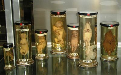
Animal Fetuses (Photo by Emily F. Porth)
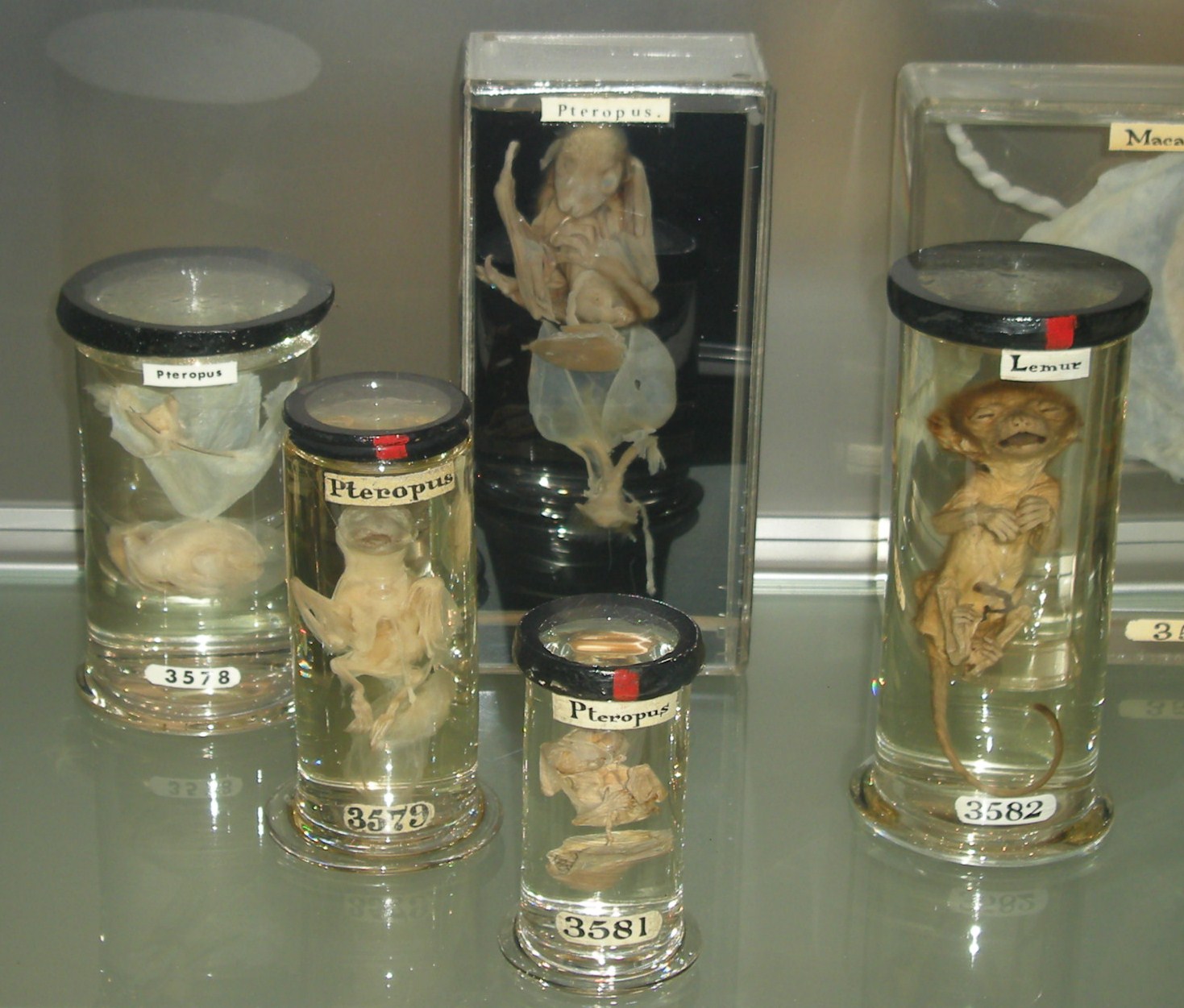
Fruit Bat Fetuses (Photo by Emily F. Porth)
Female humans and animals are marginalized or made invisible in both natural history and evolutionary exhibits, as they are frequently displayed only in the context of their capacity to bear and rear offspring (i.e., Machin; Linke). This is evident, for example, in the way museums and other popular media illustrate hominid evolution by showing only males evolving from humanity’s pre-hominid ancestors into modern homo sapiens. Females may be included as part of fetal development displays, but adult women are generally absent from the evolutionary displays that grace natural history museum galleries, except when they are depicted as “barefoot and pregnant” in the cave diorama, seated next to the fire that symbolically separates early humans from other animals.
The plastinated bodies on display in the phenomenally popular Body Worlds exhibitions are similarly problematic in their depictions of females. These exhibitions contain fetuses as well, although, unlike the Hunterian Museum, they are kept in a separate curtained area that is optional for visitors to walk through. Walter has observed that the majority of full-body female specimens (two of three in the specific Body Worlds exhibition studied) focus on the pregnant body, “implicitly defining the female as a reproductive machine” (283). Linke also documented that, while several sets of male genitalia (often elongated and erect) were displayed in Body Worlds in separate showcases, women’s reproductive organs were on exhibit with “malignancies, tumours, deformities and cysts” (17). The Hunterian also contains a large collection of penises, most of those on display from men afflicted with syphilis, but there is little to illustrate their internal reproductive organs. In contrast, at both the Hunterian Museum and in Body Worlds, the internal reproductive organs of females are displayed, rather than external genitalia. Perhaps one can infer from this that women’s reproductive organs are solely for the “business” of producing offspring, whereas male organs are for pleasure (although, by showing diseased penises at the Hunterian, there is clearly a cautionary message as well). Once again, this type of anatomical display works to confine being female to the sole purpose of bearing offspring, and it also confirms destructive gender stereotypes in Western society.
These types of attitudes towards females did exist socially in the past, before the embryological worldview became dominant – women who chose not to marry, or women who could not bear children, were often marginalised or treated as less valuable and inferior. While women today are still judged by whether or not they choose to bear or raise children, they are also valued for the many other skills and talents they bring to society. These other contributions outside of reproduction are not, however, represented in scientific displays, and females of all species continue to be exhibited – and sometimes treated in life — almost exclusively as reproductive beings.
Narrating Fetal Remains
Although display labels providing a narrative about the specimens are few and far between in the Hunterian, when a story is conveyed in the museum gallery it is critical to consider whether the narrative has been misrepresented by being re-interpreted through the embryological origin story. In one example from the Hunterian Museum, a display label describes a tissue specimen (3590) consisting of:
the left ovary, fallopian tube, and half of a uterus from a woman in the first month of pregnancy… These specimens come from a woman aged 25. She was a domestic servant who committed suicide by taking poison. According to John Hunter, who carried out the post-mortem examination, she was pregnant, although her family suggested that her last period had been less than one month before. At the time it was suggested that the lateness of her period and the realisation that she was pregnant may have prompted her to take her own life. (RCSENG 2011)
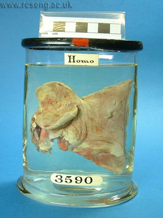
Specimen 3590 (Photo courtesy of Royal College of Surgeons)
The name of the woman was Mary Hunt, and she died on April 20, 1792. Given the previously discussed late eighteenth century views on pregnancy, it seems likely that the speculative story presented about her condition, and her reaction to it, may actually be quite fictitious. In this account by the museum, the voices of the woman and her family are silenced to privilege the perspective of the male surgeon. If she had not yet even missed her period, one wonders about the circumstances surrounding her impregnation, which are not at all investigated or considered; being in a subservient position in a wealthy household, within a society that was remarkably stratified along the lines of class and gender, it is possible that she experienced some sort of sexual violence which led to her pregnancy. It is doubtful that the attending doctor would have questioned such circumstances, particularly if it reflected poorly on her employer, who was likely an acquaintance or friend of his. No one has questioned whether she committed suicide because of the situation which led to her pregnancy, rather than the pregnancy itself, which she was likely not yet aware of — if Hunter was correct, and she was, indeed, pregnant. It can be said that this alternative narrative is just as unlikely or problematic as the one the museum tells, but that is exactly the point.
What is further concerning is the way that an artist, Karen Ingham, chose to give Mary a voice through an exhibition she created at the Hunterian in 2009 called “Narrative Remains.” Ingham chose six specimens from the Hunterian collection and, using the information from the museum database, she constructed first person narratives about each of these specimens – voices that spoke from beyond the grave. She imagines Mary would now say to an audience:
I didn’t want to do it but once my employer knew I was pregnant I’d be out on the street and as good as dead anyway. There was nothing for it, not for the likes of a poor girl like me. The poison did its job and I thought that was the end of it but the surgeon John Hunter thought different. He opened me up and poked here and there exclaiming this way and that with fancy names in Latin I couldn’t make head nor tail of. But he seemed to find me of interest, which was gratifying, for gentlemen of his class never found me of interest when I was alive. They’d look right through me, which is funny really, as that’s what you’re doing now, looking through the bits of me, here in this glass jar. Looking where the baby was, too small to really see or even feel, but he knew just what had come to pass. And now here I am, in a place with other bits of mothers and babies, so it’s almost like I’m part of a family after all. (Ingham)
In this narrative, Mary appears gratified and honored that she has been dissected and put into a jar by Hunter, suggesting that women through history have wanted nothing more in life than to have the attention of a man, particularly if he is wealthy – even if they have to die for it. Ingham also seems to equate being from a lower class with being unaided and emotionally destitute, as she implies Mary has no family (aside from preserved bits of mothers and babies) and thus no options for survival if she were to lose her job. However, based on the account in the museum catalogue of her family’s testimony, which indicates they shared a close enough relationship to know her menstrual cycle, it seems highly unlikely that she was uncared for and alone in her life. Finally, the narrative exalts Hunter’s knowledge and expertise in knowing “just what had come to pass,” when he clearly did not know any more about the situation beyond that it was likely she had conceived.
The Question of Context
My position is that museums should provide a context about their specimens in order to honor the former lives of those beings, as well as to aid visitors in connecting to and learning from them. Yet, as the story told about Mary Hunt demonstrates, narratives become dangerous when they translate the past through a contemporary worldview that is contextually insensitive. This is especially true when such stories contain ideas that actually serve to reinforce harmful stereotypes about women in contemporary society.
As with human specimens, I believe it is also possible to enable a meaningful connection between visitors and nonhuman animal specimens by situating the latter within a context. Some of the display labels that accompany animal specimens in the Museum illustrate the context of their procurement, but most do not. In the case of some specimens, labels clearly indicate that the animal was killed specifically for research purposes, or to obtain its offspring. For example, the label on a jar containing a secretion from the uterus of a sheep (specimen 3487) notes that the animal was killed three days after copulation specifically “to show changes in the reproductive organs of ewes in the days following impregnation” (RCSENG 2011). The detailed labelling seems to be most common among the domesticated animals that Hunter raised himself, whereas the more exotic animal specimens tend to have labels even more ambiguous than the human specimens: for example, “A fetal infant armadillo near the end of its gestation”(specimen 3479).
One exception is a rhesus monkey fetal specimen (specimen 3585), which is described in the catalogue as having “come from a female monkey owned by a 'Mr Endersbay' (probably William Endersby of Bedfordshire, d.1802)… The monkey gave birth on 15 December 1782, and the specimen would have been prepared shortly afterwards” (RCSENG 2011). There is no indication of whether the monkey was stillborn or killed at birth for Hunter’s collection, and the sex remains unmentioned, even though it is clearly male from the photograph in the catalogue. Further, there is no reference to the well-being or circumstances of Mr. Endersby’s rhesus monkey after she mothered the specimen. The lack of details makes it difficult to feel any sort of empathy or connection to the monkey on display or to his mother, which could assist visitors in learning about the role that animals played in relation to the history of surgery, and about colonialism and the trade in exotic animals. It could also prompt museum audiences to reflect on how the contemporary exotic pet industry remains problematic.

Specimen 3585 (Photo courtesy of Royal College of Surgeons)
The visitors I interviewed were divided about whether they wanted more descriptive labels in the Museum in order to situate the remains within a context. John commented that he was more bothered by looking at the surgical instruments than at the fetal specimens, “because it’s easy to imagine what they did with them after reading the stories!” (18/06/10). Penny also reinforced this by saying she had a “sympathetic pain reaction” after learning about surgery in the eighteenth century and seeing the size of gallstones and the tools used to remove them; she felt pity for the people represented in the museum because they were “the worst cases” (16/06/10). Interestingly, these stories help visitors connect with and understand what the experience of surgery would have been like in a historic context, yet in general they do not seem to help visitors to understand the lives of individuals whose anatomical contributions generated new understandings of the body and disease, and who were a part of how the field progressed to make surgery safer for others.
Alexa thought it was better not to have the histories of the human fetal specimens included because “people might have a strong emotional reaction to the fetuses if their stories were known — but it doesn’t have to be a bad reaction, either!” (25/06/10). Anne (24/06/10), a “slightly spiritual” British woman working on her psychology undergraduate degree, was concerned that, by putting more emphasis on who the specimen was in life, the Museum also “muddies the ethical waters about their responsibility to their visitors” in terms of their mental health. To situate her response within a context, Anne told me she had been one of three triplets, but her “other sisters didn’t make it… it was not hard to see [the fetal bodies], I was just curious, but my mum wouldn’t have been able to see them” (24/06/10). It seems as though her concerns about the Museum’s responsibilities to the mental health of its visitors may be valid in this case, although all of the people I spoke with seemed quite prepared for the type of specimens they might encounter because of the nature of the Museum.
Females as the “Absent Referent”
By removing females completely from the embryological origin story and highlighting the value of the fetus, but not the value of females who bore them, the latter are effectively rendered symbolically worthless. This has serious repercussions for the lives of females, especially when the well-being of the unborn is privileged over the well-being of the living. Females have become the “absent referent,” in the way that Carol Adams uses the term to refer to the psycho-social detachment experienced by people who eat meat. It explains how there is a total disconnect between living animals and the end product that ends up at the dinner table:
One does not eat meat without the death of an animal. Live animals are thus the absent referents in the concept of meat. The absent referent permits us to forget about the animal as an independent entity; it also enables us to resist efforts to make animals present. The absent referent functions to cloak the violence inherent to meat eating, to protect the conscience of the meat eater and render the idea of individual animals as immaterial to anyone’s selfish desires. The function of the absent referent is to allow for the moral abandonment of a being. (Adams)
When gazing at the fetus as it has traditionally been displayed, females are the absent referent. “Most of the fetuses do not even have an umbilical cord, a memory to a maternal relationship. They appear out of context to a woman, to flesh, to a placenta, to origin… their author is the anatomist” (Duden 23). Fetuses are presented almost as though they were immaculately conceived – but without the Virgin Mary. Most of my interviewees said they were more affected by seeing wet-preserved fetal bodies than by seeing the fetal skeletons, and Amir phrased the difference in these terms: “because it was in fluid, it was striking because it looked so much like it would in the womb, but with a man-made jar around it, the analogy is quite disturbing… the effect of the glass and water magnifies it too, enhancing the impact because of the detail” (21/06/10). The jar is a glass womb, a male uterus. It is a very literal metaphor in that it appears innocuous, but the jar is actually the absent referent for the male surgeon-anatomist who wanted to “give birth” to the fetus that he collected and then re-animated. It is the referent which obscures the absent referent through its transparency. In a way, it justifies fetal displays because it allows them to occur; its message is that a female is not needed, and it posits the male womb as an objective space that the female womb can never hope to be. Through its transparency, it claims that context is not required.
Fetus in Amnion (Photo by Emily F. Porth)
As the absent referent in fetal display, females are effectively morally abandoned as “incubators.” That this has become a lived reality for some beings is especially true of domesticated female animals in factory farm industries, who are valuable only as long as they can reproduce, regardless of how desperate the condition of their overall health, or how much emotional stress they experience. They are discarded as waste after becoming “spent” and losing their ability to reproduce. The industrial farming of animals came into being with chickens in the early twentieth century (Boyd), and although this was well after the embryological worldview had been established, it can be said that this system of practice only became possible because females had already been morally abandoned.
Given the autonomy of chicken embryogenesis and the increased availability of reliable energy in the form of electricity, thermostatically regulated incubators substituted for the brooding hen, allowing large numbers of newly hatched chicks to be produced on demand… By 1934, roughly half of all chickens raised in the United States were hatched in artificial incubators, with state of-the-art hatcheries operating as "veritable chick factories" capable of producing more than one million chicks per year. (Boyd 639-640)
It is important to note that this type of agriculture is destructive for males too, whether they are male calves deemed useless by the dairy industry and then confined to a dark crates for the rest of their short lives (Fudge 38), or the innumerable male chicks who are destroyed almost immediately after hatching in the egg production industry (Cassuto 64).
The agricultural industrial complex is an institution that completely precludes females from rearing their young (and, in the case of the egg industry, even from the opportunity to bring them into the world), and only values them as incubators of a “product.” This system has, however, only been able to come into existence because the embryological worldview has symbolically rendered females as marginal to the production of her offspring.
Reimagining an Inclusive Fetal Display
The issue of whether fetal remains should be on display at all is contentious, particularly when images of the dead fetus have been used in a way that can be described as violent by anti-choice groups in relation to the abortion debate. Yet, this is precisely why Morgan advocates for continued exhibition of fetal displays: “as long as fetal remains appear only to symbolize abortion, this link will remain unchallenged… the less that fetal remains are visible in public, and the more limited the contexts in which they appear, the more shocking and sordid their appearance comes to seem” (31). Morgan, however, does little to further a new vision of how fetal remains could be displayed differently to challenge the assumptions of the embryological origin story. I ask two questions in my quest to reimagine fetal display: first, how can museums better represent the lives of females in scientific displays, honoring them as mothers and yet also for the many other things they do? And second, how can museums incorporate other worldviews about human relationships with animals that encourage attitudes of respect and kinship?
One of my interview questions challenged visitors by explaining that some museums have been critiqued for showing females solely in regard to their reproductive capacity, while using male bodies as the standard frame of reference; I then asked whether they thought the Hunterian Museum specimen displays followed the same trajectory. While most interviewees first responded to the question with “I’d never thought of that before!,” upon reflection many of them did conclude that the Hunterian replicates this division in its displays. However, it was interesting that the visitors I spoke with thought this was allowable because of the museum’s historic context — they assumed that, for Hunter, the male body would have been the biological norm, and females would have been valued primarily in terms of their reproduction, and if that is how he did it, that is how the specimens should be displayed. This is interesting, and it could potentially complicate attempts by the Museum to make its displays more inclusive, lest they be judged as “inauthentic.” As such, it is special exhibitions and other supplementary materials that may be the best way for the Hunterian and other medical museums to challenge the embryological origin story and incorporate other ways of knowing.
At the Hunterian Museum, females associated with the collections can regain their visibility through changes to both the museum guide and the audio guide. Although little has been recorded about the origins of the fetal remains in Hunter’s collection, a historical context could be provided that discusses how women at the time would have understood pregnancy and the child growing within them. Ultimately, if museums are going to display fetal remains, especially those that were collected historically, they need to be transparent about changing perceptions of the fetus because contemporary audiences will interpret them so differently. Females could also be given a more audible and contemporary voice by incorporating narratives from individual women about experiencing the loss of a pregnancy, or by using stories from zoologists and other animal professionals (from zoos, farms, game parks, etc.) who have an understanding of non-human animal behavior in response to pregnancy and to the death of a child. These voice recordings could then be featured as part of the museum audio tour, and help audiences to have an empathetic reaction and emotional connection to the specimens they are viewing. Additionally, females are frequently focused on as “mothers” in museums, yet exhibitions seem largely to disregard the role that males play as fathers (Machin 60). As such, it would be complementary and interesting to provide information about the changing role of men within the domestic sphere during this period, and whether their understandings of reproduction changed in the same ways and at the same times as did women’s.
The Hunterian Museum also offers daily volunteer-led tours that center on different themes, and a tour focusing on the daily life of women in the 18th century, situated within the contexts of class and ancestry, from motherhood and beyond the domestic sphere, would be an excellent way to contextualize the fetal bodies and other remains of women in the galleries. The museum could also feature a tour specifically about the lives of animals in the 18th century and their various relationships with humans, as it would situate the non-human animal remains (and particularly the circumstances of their acquisition) within the context of colonialism and changing scientific and religious worldviews. It would also help to make the lives of animals more significant to visitors. Additional information, where known, could be provided in the Hunterian Museum’s online catalogue; although I was told there is no further data available about the context of the human fetal remains in Hunter’s collection, basic details such as the sex of animal fetal bodies could easily be added. This is a very simple way to acknowledge the animals as individuals and recognize females in the collection.
Special exhibitions at the Museum have an even greater potential to provide insight into other origin stories and ways of knowing, and the Hunterian Museum has a strong history of these which can both supplement and challenge the permanent displays.15 One of the main assumptions that a special exhibition could defy is the idea that humans experience both physical and social evolution, but non-human animals do not: animal and human fetuses could be displayed side by side at equivalent stages of development, accompanied by an explicit discussion about human-animal evolutionary kinship. This could be supplemented by information about the changing relationships between humans and other animals from the eighteenth century. Also, numerous specimens of the same animal species could be shown together at different stages of development, mirroring the way that human fetal specimens are traditionally displayed. These display techniques would reflect a worldview in which animals and humans are kin and humans are not perceived to be special or superior to other animals. To take this exhibition one step further and challenge the embryological origin story, the museum could include origin stories that do not consider a fetus to be a biological reality – this could be within a historical context, or narrate the contemporary beliefs of certain religious or cultural groups who hold a different idea of what a fetus is. Morgan documents, for instance, how some women in Ecuador today do not think of a fetus or young baby as a person — it is “more like a puppy, a little animal.” Even though they practice Catholicism, it is not quite a person until it is baptized (xii-xiii).
A special exhibition would also be the ideal place to draw links between historical bodies and contemporary issues. For example, an exhibition could focus on the way female animals were, and continue to be, exploited for their reproductive capacity by reflexively discussing the way female bodies are displayed in the museum, and how that reflects the way Western society uses and values female animals. Narratives could describe first-person accounts of respectful human-animal interactions and relationships in different contexts, from various cultures or faith traditions and through different periods of time. For example, this could include narratives from Christians who describe their beliefs about the role humans play in acting as God’s stewards of the natural world. Through these sorts of alternative interpretations, even anatomical museums can pose challenging questions to visitors that have the potential to result in positive social change.
Finally, the Museum could also take steps to temporarily alter its permanent galleries. For instance, Machin documents how the Manchester Museum drew attention to the invisibility of female animals in its natural history galleries by enacting an “intervention.” Staged during International Women’s Week (March 4th to 11th), the intervention involved placing a single female specimen prominently in the central aisle of the mammal gallery as an introduction to the initiative (57), and then concealing male taxidermy specimens with white sheets to draw attention to the insignificant number of specimens representing females (59).16 A small display label accompanied the concealed specimens, explaining the intervention and describing the female specimens that were still in storage in order to highlight the inadequacy of female representation (59).
This example is important because of the tendency within science to describe females as different from males, “implying that the male state represents the standard from which females deviate” (Machin 57). In a museum like the Hunterian that is full of anatomical specimens – just bits and pieces of bodies – it is usually impossible to determine the sex of the individual who contributed the specimen unless it is explicitly noted on the display label, and often it is not. Hence, visitors assume the specimens are from male bodies, unless the biological bits deal specifically with the reproductive system and are thus easily identifiable. As Machin eloquently states,
if museum visitors are to see animal diversity properly represented, one might expect to find that female and male animals are proportionately shown in museums… there is a curatorial obligation or responsibility to explain the collections on display and to encourage visitors to reflect on the extent to which displays properly represent difference and diversity with respect to life on earth. (55)
It is this “curatorial obligation” to “represent difference and diversity” that all museums, even those purporting to “objectively” display their scientific specimens, need to consider.
Concluding Thoughts
Museums, particularly those exhibiting scientific collections, may be hesitant to make their displays more socially inclusive by deconstructing their exhibitions and incorporating other perspectives. However, as I have demonstrated through an examination of the way fetal specimens are displayed at the Hunterian Museum, such scientific exhibitions are loaded with cultural assumptions that prove to be harmful to females of all species. This is characterized by the way females have become the “absent referent” in regard to fetal remains, and the lack of contextualization regarding the changing understanding female bodies and of pregnancy from the eighteenth century. This multifaceted “hidden curriculum” embedded within the fetal displays at the Hunterian Museum and in other similar exhibitions ultimately reinforces the marginalization and exploitation of females in Western society.
Museums can promote inclusivity at three levels — individual, community, and society (Dodd and Sandell 25) — and it is on a societal level, on which museums can “contribute towards the creation of more equitable societies” that I suggest the Hunterian Museum can make changes to help visitors move beyond the sexist and speciesist notions that are embedded within scientific displays of human and animal bodies. Museums need to go beyond deconstructing exhibitions and instead commit to re-imagining and enacting inclusive exhibitory techniques for these same displays. By providing the suggestions outlined above, it is my hope that museums will incorporate other ways of knowing that challenge the embryological origin story, and also re-position females of all species in ways that enable visitors to identify with them, thereby encouraging respect and learning. It is not only necessary to provide females with adequate physical representation in museum displays, but also to allow specimens to speak with voices that are situated within the correct historical context. The authoritative voice of the white male surgeon needs to be de-centered in order to allow for this to happen. As Smith eloquently states:
How Aristotle departed from Plato, what Stalin said to Churchill, and how Emerson engaged with Thoreau - these and like topics consumed my class time. Scholarly fields ranging from history to philosophy to science, literature, and law have developed through the medium of male conversation. If I have benefited from this listening, might not men... learn from listening to a conversation among women? (67)
Finally, it is important to emphasize that it is not enough to deconstruct and reimagine museum displays from a feminist perspective, if that perspective remains anthropocentric. As I have demonstrated, there are innumerable parallels between the ways that females of every species can be exploited for their reproductive capacity, and all are impacted through the widespread acceptance of the embryological origin story and the problematic ways it is popularly depicted. It is only through recognising these similarities, and in accepting that various forms of oppression are interlocking (Plumwood), that real change can be made.
Notes
1. It should be noted that the present day catalogue entry describes this kangaroo as an “infant” or “newborn,” rather than as a fetus. Marsupials are born before they have fully developed to survive outside of their mother, and instead complete their development inside their mother’s pouch. The described kangaroo specimen is still too young to have survived outside of the pouch and, as such, can be similarly described and compared to other fetal animals.
2. All interviews were semi-structured and open-ended, and coded manually for analysis. All names used are pseudonyms, unless individuals requested otherwise.
3. One of my interviewees referred to the fetal specimens as “stillborns,” as he assumed that this was the natural circumstance which would have brought them to the Hunterian Museum (Luke 24/06/10). This is interesting, especially because the museum is somewhat transparent in the exhibition text about the strong likelihood Hunter acquired bodies from grave diggers. However, most of the people I interviewed made no such assumptions, and he seems to be an exception.
4. In relation to the biological development of a human being, the term “embryo” refers to the being from fertilization to the seventh week of gestation. From week eight until birth, the being is referred to as a “fetus,” the idea being that it can be described as such when it begins to look like the species to which it belongs (Morgan 91). I use the term fetus because I am dealing with museum specimens that are generally further along in their development and easily recognizable as belonging to a particular species.
5. This paper is not about the abortion debate, but it is important to acknowledge that the anti-choice movement has done an excellent job, particularly in North America, of associating fetal bodies with terminated pregnancies. I think it is important for the public and average layperson to see fetal bodies in a context like a museum that is without stigma in order to dissociate them from the abortion debate.
6. It is important to note that not all human bodies have been displayed solely in cultural history institutions. There is a tragic history of indigenous peoples being displayed, both living and dead, at natural history museums and zoological parks. Ota Benga, for instance, was a living Congolese man brought to the United States who was first exhibited at the St. Louis World’s Fair in 1904, and then as a living exhibit at the American Museum of Natural History in New York. Benga “failed to be sufficiently docile in the museum,” and so he was contracted out to the New York Zoological Gardens where he was put on display in the primate house; his exhibition was “a huge success,” although he was eventually released into the care of clergy after advocacy groups intervened to protest his display as inhumane (Ota Benga Alliance).
7. There is another Hunterian Museum, featuring the collection of William Hunter, at the University of Glasgow in Scotland.
8. Owen, while probably best known for coining the term “dinosauria,” went on to found the Natural History Museum in London (Fortey 37).
9. Teratology literally means “the study of monsters” (Asma 66) and refers to biological organisms with developmental anomalies. As Anker and Franklin note, “much of the ‘stilled’ life packed into ‘wet’ tissue collections was ‘failed’ in the sense of displaying pathological disease, deformity, and death, illustrating, among other things, that life was both radically plastic and capable of veering ‘off plan’” (105). The fetal specimens I focus on in this paper that are on display to the public at the Hunterian Museum are not, however, obviously deformed or otherwise “monstrous.”
10. I, and many others, do not agree that evolution is necessarily devoid of divine energy or intent. Evolution and religion have, however, been popularly portrayed as being polarized, particularly in a North American context, and I do not wish to represent them as such in my research.
11. The label, however, does not say that the mother of the babies was named Margaret Waddington, born in 1765. She was twenty-one when she gave birth to the quintuplets, and already had one older child as well (RCSENG 2011). The museum label focuses on the activities of the male doctor, rather than on the woman who bore them. It would have been easy to incorporate the interesting narrative about the value of the placenta compared to that of the remains, thereby creating a version of the story where the mother is a central actor in the existence of her own children.
12. The assumption that nonhuman animals will only enact heterosexual couplings, and that only two genders exist in all other species (Bagemihl), is still commonly depicted in natural history museums (Levin).
13. Readers may have noticed by this point that the majority of my interviewees were all highly educated. Given that interviewees were approached completely randomly in the Museum, it seems apparent that it attracts a certain type of visitor because of the nature of its collections and its affiliation with the Royal College of Surgeons.
14. There were a few exceptions to this in the Hunterian Museum, including the kangaroo fetal specimens.
15. Ingham’s Narrative Remains was one of these special exhibitions. Although the fictionalized stories it puts forth are highly problematic, the premise of the exhibition is quite excellent and it was well-presented in a way that was very engaging.
16. In the Manchester Museum natural history galleries, only 11% of specimens were represented by females alone, and 14% of species were represented by both male and females specimens (Machin 58).
Works Cited
Adams, Carol J. “A vegan-feminist lament.” [2009] Carol J. Adams (blog), 31 July 2011.
Adams, Carol J. and Josephine Donovan. “Introduction.” Animals and Women: Feminist Theoretical Explorations. Carol J. Adams and Josephine Donovan, Ed. Durham, NC: Duke UP, 1995. 1-8.
Anker, Suzanne and Sarah Franklin. “Specimens as Spectacles: Reframing Fetal Remains.” Social Text 106, 29.1 (Spring, 2011): 103-125.
Asma, Stephen T. Stuffed Animals and Pickled Heads: The Culture and Evolution of Natural History Museums. Oxford: Oxford UP, 2003.
Bagemihl, Bruce. Biological Exuberance: Animal Homosexuality and Natural Diversity. New York: St. Martin’s, 1999.
Boyd, William. “Making Meat: Science, Technology, and American Poultry Production.” Technology and Culture 42.4 (2001): 631-644.
Buklijas, Tatjana and Nick Hopwood. 2010. Making Visible Embryos. Accessed July 19, 2011.
Campbell, Sue, Letitia Maynell, and Susan Sherwin. “Introduction: Minding Bodies.” Embodiment and Agency. Sue Campbell, Letitia Maynell, and Susan Sherwin, Ed. College Station: Pennsylvania State UP, 2009. 1-21.
Cassuto, David N. “Bred Meat: The Cultural Foundation of the Factory Farm.” Law and Contemporary Problems 70.59 (Winter, 2007). 59-87.
Christian, William A. Visionaries: The Spanish Republic and the Reign of Christ. Princeton, NJ: Princeton UP, 1996.
Coates, Peter. Nature: Western Attitudes Since Ancient Times. Los Angeles: U California P, 1998.
Darwin, Charles. On the Origin of Species by Means of Natural Selection, or the Preservation of Favoured Races in the Struggle for Life. London: John Murray, Albemarle- Street, 1859. http://darwin-online.org.uk/pdf/1859_Origin_F373.pdf, accessed May 9, 2012.
Desmond, Jane. “Postmortem Exhibitions: Taxidermied Animals and Plastinated Corpses in the Theaters of the Dead.” Configurations 16.3. (Fall 2008): 347-377.
Dobson, Jessie. “RCSHC/3773 – Macropus.” Royal College of Surgeons of England online database of museum and archive collections, 2011 [1970]. 20 May 2011.
Dodd, J. and Richard Sandell. Including Museums: Perspectives on Museums, Galleries and Social Inclusion. Leicester: Research Centre for Museums and Galleries, University of Leicester, 2001.
Duden, Barbara. “The Fetus on the ‘Farther Shore’: Toward a History of the Unborn”, Fetal Subjects, Feminist Positions. Lynn M. Morgan and Meredith W. Michaels, Ed. Philadelphia: U Pennsylvania P, 1999. 13-25.
Foltyn, J.L. “The corpse in contemporary culture: Identifying, transacting, and recording the dead body in the twenty-first century.” Mortality 13.2 (May 2008): 99-104.
Fortey, Richard. The Secret Life of the Natural History Museum: Dry Store Room No. 1. London: Harper Perennial, 2008.
Franklin, Sarah. “Dead Embryos: Feminism in Suspension.” Fetal Subjects, Feminist Positions. Lynn M. Morgan and Meredith W. Michaels, Ed. Philadelphia: U Pennsylvania P, 1999. 61-82.
Haraway, Donna J. Primate Visions: Gender, Race, and Nature in the World of Modern Science. New York: Routledge, 1989.
Hooper-Greenhill, Eilean. Museums and the Interpretation of Visual Culture. London: Routledge, 2000.
Ingham, Karen. Narrative Remains. Wellcome Trust and The Hunterian Museum, 2009.
Jordanova, Ludmilla. “Interrogating the Concept of Reproduction in the Eighteenth Century.” Conceiving the New World Order: The Global Politics of Reproduction, Faye D. Ginsburg and Rayna Rapp, Ed. Berkeley: U California P, 1995. 369-386.
Kinsley, David. “Christianity as Ecologically Harmful” and “Christianity as Ecologically Responsible.” Ecology and Religion: Ecological Spirituality in Cross-Cultural Perspective. Englewood Cliffs: Prentice-Hall, 1995. 103-24.
Levin, Amy. “Introduction.” Gender, Sexuality, and Museums: A Routledge Reader. Amy K. Levin, Ed. New York: Routledge, 2010. 1-11.
Linke, Uli. “Touching the corpse: The unmaking of memory in the body museum.” Anthropology Today 21.5 (October 2005): 13-19.
Machin, Rebecca. “Gender representation in the natural history museum galleries at the Manchester Museum.” museum and society 6.1 (2008): 54-67.
Martin, Emily. “The Egg and the Sperm: How Science Has Constructed a Romance Based on Stereotypical Male-Female Roles.” Signs 16.3 (Spring 1991): 485-501.
Martin, Emily. The Woman in the Body. Boston: Beacon Press, 1987.
Morgan, Lynn M. Icons of Life: A Cultural History of Human Embryos. Berkeley: U California P, 2009.
Morgan, Lynn M. and Meredith W. Michaels. “Introduction.” Fetal Subjects, Feminist Positions. Lynn M. Morgan and Meredith W. Michaels, Ed.Philadelphia: U Pennsylvania P, 1999. 1-9.
Newman, Karen. Fetal Positions: Individualism, Science, Visuality. Stanford: Stanford UP, 1996.
Noske, Barbara. Humans and Other Animals: Beyond the Boundaries of Anthropology. London: Pluto Press, 1989.
Ota Benga Alliance. “Who Is Ota Benga?” 2010. 5 March 2010.
Plumwood, Val. Feminism and the Mastery of Nature. London: Routledge, 1993.
Preece, Rod. Animals and Nature: Cultural Myths, Cultural Realities. Vancouver: UBC Press, 1999.
Preece, Rod. “Darwinism, Christianity, and the Great Vivisection Debate.” Journal of the History of Ideas 64.3 (2003): 399-419.
RCSENG. SurgiCat: the online catalogue of the museum and archive collections at the Royal College of Surgeons of England, 2011. 19 July , 2011.
Sandell, Richard (Ed.) Museums, Society, Inequality. Routledge: London, 2002.
Schiebinger, Londa. Nature’s Body: Gender in the Making of Modern Science. [1993] 2nd edition. New Brunswick NJ: Rutgers UP, 2004.
Schiebinger, Londa. “Feminist History of Colonial Science.” Hypatia 19.1 (Winter 2004): 233-254.
Schiebinger, Londa. “Introduction.” Feminism and the Body. Londa Schiebinger, Ed.New York: Oxford UP, 2000. 1-24.
Smith, Barbara Clark. “A Woman’s Audience: A Case of Applied Feminist Theories.” Gender, Sexuality, and Museums: A Routledge Reader, Amy K. Levin, Ed. New York: Routledge, 2010. 65-70.
Walter, Tony. “Body Worlds: clinical detachment and anatomical awe.” Sociology of Health and Illness 26.4 (May 2004): 464-488.